Nottingham & Singapore Breast Pathology Online Masterclass
Advances in Breast Pathology Diagnosis and Classification – Part 2Slide Cases
The Online Masterclass includes a slide seminar session.
Each of the eight cases below will be examined and discussed in detail. We invite you to review each case and submit your own diagnosis from the choice provided before the masterclass.

For each case, we have provided the relevant images, clinical data and a number of options for the diagnosis for you to choose from. Please complete the slide survey – use the Mentimeter form to enter your diagnosis, linked HERE and at foot of this page.
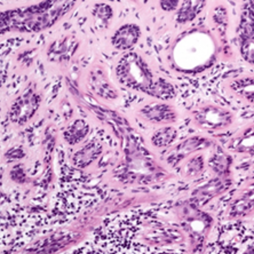
Please review the cases and submit your opinion by 5pm (UK time), 21 September.
Submitted responses will be used to guide the discussions in the case presentations. Respondent details will not be used or referred to.
Please keep a note of your choices as you choose them if you wish to refer to these during the slide seminars. Unfortunately we cannot provide a summary report of your submission.
To view the slide images
To open the image(s) for each case you will firstly need to log into the PathXL slide viewer where the images are hosted:
1. Enter the url in your internet browser: https://pathxl.co.uk
2. Against Account Name, enter: nottinghamuninhs
3. Against Username, enter: Masterclass2025
4. Against Password, enter: ERPhT2025
5. Keep the homepage open on your browser and return to the slide survey.
Now you can complete the slide survey in the Mentimeter form
If you have any issues viewing the cases, please firstly try changing your browser. For any other queries, please contact Jenny Baldwin at jennifer.baldwin@nottingham.ac.uk
Slide Seminar

Professor Puay Hoon Tan
Professor Tan is a pathologist at Luma Women’s Imaging/Luma Medical Centre. She has active interests in breast, urologic and renal pathology.
SINGAPORE
Case 1
Clinical Data:
51 year old female. Previous diagnosis of left breast cancer, ER/PR positive and HER2 negative. Current presentation of an enlarged left axillary lymph node measuring 4cm. Biopsy of the axillary mass.
Diagnosis Options:
- Metastatic ductal carcinoma
- Metastatic small cell neuroendocrine carcinoma
- Metastatic lobular carcinoma
- Metastatic lobular and small cell neuroendocrine carcinoma


Case 1 – Synaptophysin:
https://pathxl.co.uk/iscope/pxlviewer/default.aspx/?resid=257863
Professor Puay Hoon Tan
Professor Tan is a pathologist at Luma Women’s Imaging/Luma Medical Centre. She has active interests in breast, urologic and renal pathology.
SINGAPORE
Case 2
Clinical Data:
61 year old female. Uniductal bloody nipple discharge. No obvious radiological lesion. Microdochectomy performed.
Diagnosis Options:
- Intraductal papilloma
- Ductal carcinoma in situ
- Solid papillary carcinoma in situ
- Invasive solid papillary carcinoma


Case 2 – Mucicarmine:
https://pathxl.co.uk/iscope/pxlviewer/default.aspx/?resid=257867

Case 2 – Synaptophysin:
https://pathxl.co.uk/iscope/pxlviewer/default.aspx/?resid=257869

Professor Janina Kulka
Professor Kulka is Professor of Pathology at Semmelweis University, Department. of Pathology, Forensic and Insurance Medicine.
BUDAPEST HUNGARY
Case 3
Clinical Data:
58 year old lady with a 13 mm and a 9 mm spiculated mass in her right breast. Her last mammography 10 years ago was negative. Her grandmother had breast cancer (TNBC) at the age of 85. Core biopsy specimen.
Diagnosis Options:
- IBC NST
- Mixed IBC NST and ILC
- Non-classical ILC (alveolar, trabecular and with tubular elements)
- IBC NST with lobular growth pattern

Professor Janina Kulka
Professor Kulka is Professor of Pathology at Semmelweis University, Department. of Pathology, Forensic and Insurance Medicine.
BUDAPEST HUNGARY
Case 4
Clinical Data:
70 year old lady with a 10 cm mass in her right breast. The whole breast is edematous, the skin of the breast is thickened. There are enlarged lymph nodes in the axilla. Her sister had breast cancer. Core biopsy specimen.
Diagnosis Options:
- Classical ILC (with signet ring cell features)
- ILC with extracellular mucin (with solid growth pattern and signet ring cell features)
- Metastatic (poorly cohesive) gastric adenocarcinoma to the breast
- Type B mucinous carcinoma

Professor Janina Kulka
Professor Kulka is Professor of Pathology at Semmelweis University, Department. of Pathology, Forensic and Insurance Medicine.
BUDAPEST HUNGARY
Case 5
Clinical Data:
77 year old lady with a 26 mm poorly echoic lesion in her right breast. No further imaging or clinical information was provided. Core biopsy specimen.
Diagnosis Options:
- Low grade adenosquamous carcinoma of the breast
- IBC NST (poorly cellular with desmoplastic stroma)
- Metastatic (lung) adenosquamous carcinoma to the breast
- Metaplastic breast carcinoma (NOS)


Professor Hannah Wen
Professor Wen is Director of the Breast Pathology Fellowship at Memorial Sloan Kettering Cancer Center in New York.
NEW YORK USA
Case 6
Clinical Data:
40-year-old woman presented with a rapidly growing mass in the left breast. Core biopsy and subsequent mastectomy revealed a high grade malignant epithelioid and spindle cell neoplasm with chondrosarcomatous and osteosarcomatous differentiation. The tumor cells are focally positive for cytokeratin AE1:AE3 and p63, negative for CK5, SOX10, desmin and myogenin.
Diagnosis Options:
- Metaplastic carcinoma
- Malignant phyllodes tumor
- Primary sarcoma

Case 6 – HE:
click on image above
Professor Hannah Wen
Professor Wen is Director of the Breast Pathology Fellowship at Memorial Sloan Kettering Cancer Center in New York.
NEW YORK USA
Case 7
Clinical Data:
50-year-old woman, with no history of breast carcinoma, presented with a left axillary mass. Core biopsy and subsequent excision showed a poorly differentiated carcinoma with squamous differentiation.
Diagnosis Options:
- Metaplastic breast carcinoma squamous cell type
- Cutaneous squamous cell carcinoma
- Metastasis carcinoma

Professor Hannah Wen
Professor Wen is Director of the Breast Pathology Fellowship at Memorial Sloan Kettering Cancer Center in New York.
NEW YORK USA
Case 8
Clinical Data:
77-year-old woman presented with a left breast mass. Left breast lumpectomy reveals a biphasic neoplasm with atypical epithelial and myoepithelial proliferation with focal infiltrative border.
Diagnosis Options:
- Metaplastic carcinoma
- Atypical adenomyoepithelioma
- Malignant adenomyoepithelioma

CPD/CME
Each masterclass is approved by the Royal College of Pathologists (8 CPD points). Approval is pending from the Singapore Medical Council (4 CME points).


• ALL registered attendees will receive a Certificate of Participation
• The Certificate enables you to claim CPD/CME points
Send a Message
Course Directors
Prof Emad Rakha and
Prof Puay Hoon Tan
Course Moderators
Dr Areeg Abbas
Course Contact
Geoff Cross