Nottingham & Singapore Breast Pathology Online Masterclass
Advances in Breast Pathology Diagnosis and Classification – Part IISlide Cases
The Online Masterclass includes a slide seminar session.
Each of the eleven cases below will be examined and discussed in detail. We invite you to review each case and submit your own diagnosis from the choice provided before the masterclass.

For each case, we have provided the relevant images, clinical data and a number of options for the diagnosis for you to choose from. Please complete the slide survey – use the Mentimeter form to enter your diagnosis, linked HERE and at foot of this page.
Please review the cases and submit your opinion by 5pm (UK time), 12 May.
Submitted responses will be used to guide the discussions in the case presentations. Respondent details will not be used or referred to.
Please keep a note of your choices as you choose them if you wish to refer to these during the slide seminars. Unfortunately we cannot provide a summary report of your submission.
To view the slide images
To open the image(s) for each case you simply click on the thumbnail image or its associated URL.
Now you can complete the slide survey in the Mentimeter form
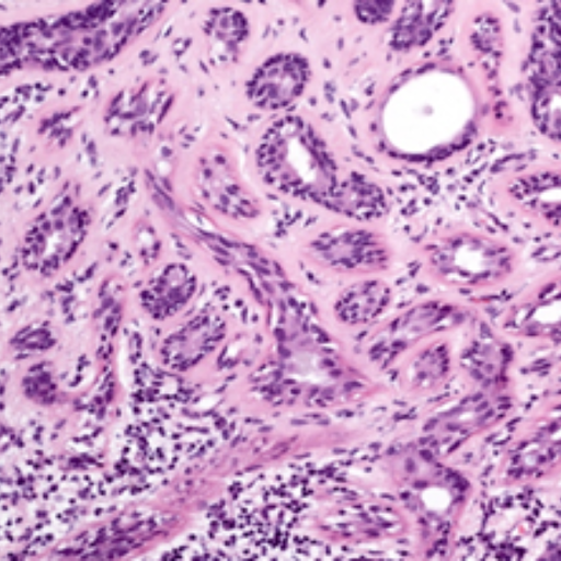
If you have any issues viewing the cases, please firstly try changing your browser. For any other queries, please contact Jenny Baldwin at jennifer.baldwin@nottingham.ac.uk
Slide Seminar


Professor Abeer Shaaban
Dr Shaaban is a specialist breast pathologist at Queen Elizabeth Hospital Birmingham and Honorary Senior Lecturer at the University of Birmingham. She trained in anatomic pathology in Liverpool, Birmingham and Leeds. She completed her PhD in molecular pathology at the University of Liverpool with the late Prof John Sloane.
BIRMINGHAM
Case 1
Clinical Data:
42 year old female. Right lower central breast mass – U3, core biopsy
Diagnosis Options:
- Papilloma with florid usual ductal hyperplasia
- Encapsulated papillary carcinoma
- Solid papillary carcinoma
- Tall cell carcinoma with reversed polarity
- Adenomyoepithelioma
NOTE: To view the images for this case, click on Case 1 – HE. When you have opened this image, view the other images by using the left hand navigation pane. Image 2 = CK5, image 3 = ER, image 4 = P63.


Case 1 – CK5:


Professor Abeer Shaaban
Dr Shaaban is a specialist breast pathologist at Queen Elizabeth Hospital Birmingham and Honorary Senior Lecturer at the University of Birmingham. She trained in anatomic pathology in Liverpool, Birmingham and Leeds. She completed her PhD in molecular pathology at the University of Liverpool with the late Prof John Sloane.
BIRMINGHAM
Case 2
Clinical Data:
90 year old female, symptomatic mass, sections from mastectomy specimen.
Diagnosis Options:
- Intraduct papilloma
- Papillary DCIS
- Serous like breast carcinoma
- Encapsulated papillary carcinoma
- Metaplastic carcinoma

Case 2 – HE (A3):

Case 2 – HE (A4):

Case 2 – P63:

Professor Hannah Wen
Professor Wen is Director of the Breast Pathology Fellowship at Memorial Sloan Kettering Cancer Center in New York.
NEW YORK USA
Case 3
Clinical Data:
60-year-old woman with right breast mass. Right breast mastectomy.
Diagnosis Options:
- Mixed mucinous carcinoma
- Invasive carcinoma, no special type, with mucin production
- Invasive lobular carcinoma, pleomorphic type, with extracellular mucin
- Mixed invasive carcinoma no special type and invasive micropapillary carcinoma

Professor Hannah Wen
Professor Wen is Director of the Breast Pathology Fellowship at Memorial Sloan Kettering Cancer Center in New York.
NEW YORK USA
Case 4
Clinical Data:
74-year-old woman with palpable left breast mass.
Diagnosis Options:
- Pure mucinous carcinoma
- Mixed mucinous carcinoma
- Invasive lobular carcinoma with extracellular mucin
- Metastatic carcinoma


Professor Edi Brogi
Professor Edi Brogi, MD PhD practices at Memorial Sloan-Kettering Cancer Center, with a focus on the diagnosis of breast diseases.
MSKCC NEW YORK
Case 5
Clinical Data:
64 year old female with breast mass. U/S guided core needle biopsy.
Diagnosis Options:
- DCIS and classic LCIS
- DCIS and florid LCIS
- (Micro) invasive lobular carcinoma, florid LCIS and DCIS
- (Micro) invasive lobular carcinoma, florid LCIS, DCIS and lymphovascular invasion (ductal carcinoma)
- (Micro) invasive lobular carcinoma, florid LCIS, DCIS and lymphovascular invasion (lobular carcinoma)


Professor Wentao Yang
Professor Yang is Professor in the Dept. of Pathology, Fudan University Shanghai Cancer Center and contributor to the 5th & 6th Edition of WHO Classification of Breast Tumours.
SHANGHAI CHINA
Case 6
Clinical Data:
48 year-old female presented with a left breast mass, measuring 2.5 × 2 × 1.5 cm, which was detected on routine physical examination.
Diagnosis Options:
- Intraductal papilloma with ADH
- Apocrine encapsulated papillary carcinoma
- Solid papillary carcinoma
- Invasive papillary carcinoma
- Invasive apocrine carcinoma

Professor Wentao Yang
Professor Yang is Professor in the Dept. of Pathology, Fudan University Shanghai Cancer Center and contributor to the 5th & 6th Edition of WHO Classification of Breast Tumours.
SHANGHAI CHINA
Case 7
Clinical Data:
Female, 62 years old, presenting with a one-month history of a palpable left breast mass.
Mammogram: A mass shadow was identified in the deep inner lower quadrant of the left breast, BI-RADS 4C.
Ultrasound: A heterogeneous lesion was observed near the inferior border of the left areola and the inner lower quadrant, measuring 41 × 14 × 18 mm. The lesion demonstrated unclear boundaries and an irregular shape.
.
Diagnosis Options:
- Metaplastic carcinoma (matrix-producing carcinoma)
- Centrally necrotizing carcinoma
- Adenoid cystic carcinoma
- Invasive breast carcinoma of no special type
- Malignant adenomyoepithelioma


Professor Gary Tse
Professor Tse is the Chairman and Professor in the Department of Anatomical and Cellular Pathology. He is also the Honorary Chief of Service of the Departments of Pathology of Alice Ho Miu Ling Nethersole Hospital and North District Hospital.
HONG KONG
Case 8
Clinical Data:
Female, 59 years old, right breast mass. Biopsy was performed and subsequently mastectomy. Sentinel lymph nodes assessment was 5 lymph node, all benign. Slide is from the mastectomy specimen.
Diagnosis Options:
- Mucinous carcinoma
- Infiltrating carcinoma with mucinous features
- Mucocele like lesion with atypical duct hyperplasia
- Fibrocystic change with florid epithelial hyperplasia
- Mucinous cystadenocarcinoma

Professor Gary Tse
Professor Tse is the Chairman and Professor in the Department of Anatomical and Cellular Pathology. He is also the Honorary Chief of Service of the Departments of Pathology of Alice Ho Miu Ling Nethersole Hospital and North District Hospital.
HONG KONG
Case 9
Clinical Data:
Female, 54. Right breast amorphous calcification on mammography in central inner breast.no mass. No architectural distortion. VAB was performed.
Diagnosis Options:
- Ductal carcinoma in situ
- Lobular carcinoma in situ
- Solid papillary carcinoma
- Florid epithelial hyperplasia
- Atypical duct hyperplasia, solid type


Professor Puay Hoon Tan
Professor Tan is a pathologist at Luma Women’s Imaging/Luma Medical Centre. She has active interests in breast, urologic and renal pathology.
SINGAPORE
Case 10
Clinical Data:
55 year old female with a 36mm mass.
Diagnosis Options:
- Cellular fibroadenoma
- Benign phyllodes tumour
- Borderline phyllodes tumour
- Malignant phyllodes tumour
- Periductal stromal tumour


Professor Emad Rakha
Professor Rakha is Professor and Honorary Consultant Pathologist, University of Nottingham and Nottingham University Hospitals NHS Trust.
NOTTINGHAM UK
Case 11
Clinical Data:
23 year old female patient, lesion. Presented with a retroareolar / 5 o’clock mass lesion measuring 24 x 15 x 16 mm in size.
Diagnosis Options:
- Atypical adenomyoepithelioma
- Malignant adenomyoepithelioma, in situ
- Malignant adenomyoepithelioma, invasive
- Salivary gland like tumour (adenoid cystic carcinoma)
- Complex sclerosing sesion involved by DCIS


CPD/CME
Each masterclass is approved by the Royal College of Pathologists (8 CPD points) plus from the Singapore Medical Council (4 CME points).


• ALL registered attendees will receive a Certificate of Participation
• The Certificate enables you to claim CPD/CME points
Send a Message
Course Directors
Prof Emad Rakha and
Prof Puay Hoon Tan
Course Moderators
Dr Areeg Abbas
Course Contact
Geoff Cross